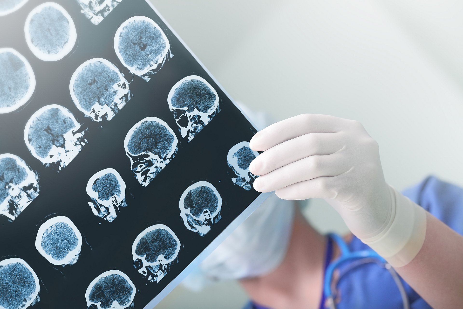
Algorithm for particularly precise assessment of brain damage
AI pinpoints stroke timing with high accuracy

A stroke occurs when blood flow to a part of the brain is obstructed, often by a blood clot. As a result, brain cells are deprived of oxygen and nutrients and die. In most strokes caused by a blood clot, medical intervention within four and a half hours can limit damage, with surgical options viable up to six hours post-stroke. As time progresses, some treatments become ineffective or may even cause additional problems
Determining the exact time of a stroke is challenging. Some strokes may start while the patient is asleep and some patients may have difficulties communicating because of the stroke symptoms. Currently, medical professionals estimate stroke timing from CT scans, where darker regions mean the stroke has progressed further. However, the unique structure of each brain makes this difficult. Even if doctors can pinpoint the approximate onset of the stroke, the individual blood flow or blood vessel structure may mean the stroke is progressing more quickly or slowly than average.
Algorithm tested on 2000 patients
Researchers from Imperial College London, the University of Edinburgh, and TUM have enhanced stroke timing estimation using artificial intelligence (AI). Trained on 800 brain scans with known stroke times, the model can independently identify affected regions in CT scans and estimate stroke timing.
Published in "NPJ Digital Medicine," the study tested the algorithm on data from almost 2,000 other patients. The software proved to be twice as accurate as using a standard visual method. The algorithm also excelled in estimating the "biological age" of brain damage, indicating how much the damage has progressed and its potential reversibility.
Enhanced precision with additional data
Leibniz Prize winner Daniel Rückert, Professor of Artificial Intelligence in Healthcare and Medicine at TUM, says: "We believe that our model is so powerful because it not only assesses how dark the damaged region is, but also includes additional features from the scans, such as texture, and accounts for variations within the damaged areas and background.”
Study leader Dr. Paul Bentley from Imperial College London says: “Having this information at their fingertips will help doctors to make emergency decisions about what treatments should be undertaken in stroke patients. Not only is our software twice as accurate at time-reading as current best practice, but it can be fully automated once a stroke becomes visible on a scan.” First author Adam Marcus even estimates that the new software could optimize treatment for up to 50 percent of stroke patients.
Marcus, A., Mair, G., Chen, L. et al. Deep learning biomarker of chronometric and biological ischemic stroke lesion age from unenhanced CT. npj Digit. Med. 7, 338 (2024). doi.org/10.1038/s41746-024-01325-z
- Original press release from Imperial College London: https://www.imperial.ac.uk/news/259073/new-ai-stroke-brain-scan-readings/
- Prof. Daniel Rückert is a member of the TUM School of Medicine and Health as well as the TUM School of Computation, Information and Technology , He is also a core member of TUM’s Munich Data Science Institute.
Technical University of Munich
Corporate Communications Center
- Paul Hellmich
- paul.hellmich@tum.de
- presse@tum.de
- Teamwebsite
Contacts to this article:
Prof. Dr. Daniel Rückert
Technical University of Munich
Chair of Artificial Intelligence in Healthcare and Medicine
+49 89 4140 8587
daniel.rueckert@tum.de

