TUM researchers combine magnetic resonance spectroscopy with fluorescence microscopy
A completely new type of microscopy based on quantum sensors

Magnetic resonance imaging (MRI) scanners are known for their ability to look deep into the human body and create images of organs and tissues. The new method, published in the journal Nature Communications, extends this technique to the realm of microscopic detail. "The quantum sensors used make it possible to convert magnetic resonance signals into optical signals. These signals are captured by a camera and displayed as images," explains Dominik Bucher, Professor for Quantum Sensing at the TUM School of Natural Sciences and researcher at the Cluster of Excellence Munich Center for Quantum Science and Technology (MCQST).
Diamond chip acts as a quantum sensor
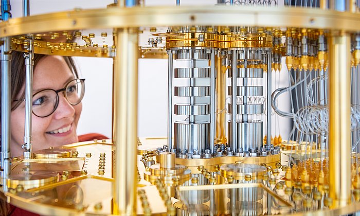
The resolution of the new MRI microscope reaches ten-millionths of a meter - that is so fine that even the structures of individual cells can be made visible in the future. At the heart of the new microscope is a tiny diamond chip. This diamond, specially prepared at the atomic level, serves as a highly sensitive quantum sensor for MRI magnetic fields. When irradiated with laser light, it generates a fluorescent signal containing the MRI signal's information. This signal is recorded with a high-speed camera and enables images with a significantly higher resolution down to the microscopic level.
A wide range of practical applications are possible
The potential applications of magnetic resonance microscopy are up-and-coming: In cancer research, individual cells could be examined in detail to gain new insights into tumor growth and spread. In pharmaceutical research, the technology could be used to efficiently test and optimize active ingredients at a molecular level. It also offers excellent potential in materials science, such as analyzing the chemical composition of thin-film materials or catalysts.
The team has applied for a patent for its development and is already planning to develop the technology further to make it even faster and more precise. In the long term, it could become a standard tool in medical diagnostics and research. "The fusion of quantum physics and imaging opens up completely new possibilities for understanding the world at the molecular level," emphasizes first author Karl D. Briegel.
Karl D. Briegel, Nick R. von Grafenstein, Julia C. Draeger, Peter Blümler, Robin D. Allert & Dominik B. Bucher: Optical widefield nuclear magnetic resonance microscopy, published in: Nature Communications, February 03, 2025, https://doi.org/10.1038/s41467-024-55003-5
- The start-up team QTAS has already been formed based on the new technology. Using magnetic resonance microscopy, it aims to detect individual cancer cells with high precision and at short intervals. The team has received the Medical Valley Award 2024 from the Bavarian Ministry of Economic Affairs.
- The research project was funded by the Bavarian State Ministry of Science and the Arts as part of the IQSense project via Munich Quantum Valley (MQV), by the Federal Ministry of Education and Research (BMBF) as part of the VIP+ Validation funding program and the Munich Center for Quantum Science and Technology (MCQST).
- Quantum science at TUM

Technical University of Munich
Corporate Communications Center
- Ulrich Meyer
- presse@tum.de
- Teamwebsite
Contacts to this article:
Prof. Dr. Dominik Bucher
Technical University of Munich (TUM)
Chair of Quantum Sensor Technology
Phone +49 89 289 13374
dominik.bucher@tum.de
https://www.ch.nat.tum.de/en/qsens/home/