Changes in fat metabolism of colorectal cancer cells demonstrated for the first time
Molecular Achilles heel of cancer cells discovered
![[Translate to en:] Darmkrebs Fettstoffwechsel](/fileadmin/_processed_/8/5/csm_210914_OM_Darmkrebs_Fettstoffwechsel_d0141c850b.jpg)
Some past measurements have also indicated significant differences in how fat is metabolized by healthy and cancerous cells. However, the results of this work have been highly inconsistent. Some investigations appeared to support such differences while others reported contrary results. “This question has been hotly debated”, says Prof. Klaus-Peter Janssen, a biologist at TUM’s university hospital Klinikum rechts der Isar.
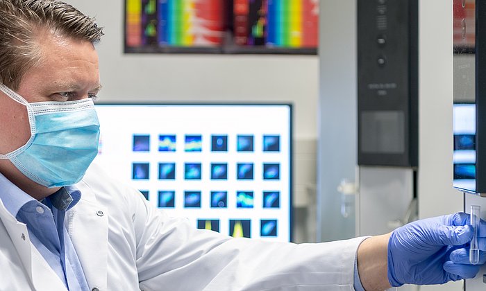
To obtain clarity, surgeons at Klinikum rechts der Isar took tissue samples from the surgically removed tumors of 144 colorectal cancer (CRC) patients. The samples were immediately prepared onsite and then analyzed using mass spectrometry at the Institute for Food & Health (ZIEL) in Freising and at the University Hospital Regensburg. This is a biochemical procedure for the quantitative measurement of the type and mass of certain molecules in specially prepared tissue samples – in this case around 200 different fat molecules.
To prove that the results were reproducible, and not merely accidental, the patients were divided into two cohorts. The tissue samples were then analyzed separately and the results compared. In addition, the researchers incorporated the analysis of samples from another cohort of 20 CRC patients investigated independently at the University of Dresden.
Like a fingerprint: Specific lipid signature demonstrated for CRC cells
In all three cohorts, the researchers were able to show that “CRC cells indeed have a specific lipid signature,” says Janssen. This means that they show a certain pattern of different lipid molecules – “A fingerprint, in a sense, with which we can distinguish between cancer cells and normal cells with a high degree of certainty. This signature also has prognostic relevance. In other words, it can be used to predict the course of the illness.”
The changes in the lipidome – i.e. the totality of lipids in a cell – related mainly to sphingo- and glycerolipids. These differences were also reflected at the genomic level: The team showed that the activity of certain genes that provide the blueprint for various enzymes was also significantly altered. Enzymes are functional proteins that play an important role in the production of metabolic products such as lipids, for example. This might be a starting point for cutting off the energy supply to cancer cells and thus slowing their growth – by finding drugs to activate or inhibit individual enzymes in order to starve the cancer.
Close cooperation crucial to successful research
“Lipids in tissue samples are highly sensitive molecules that are sometimes subject to rapid change or decay,” says Janssen. Unless tissue samples are shock-frozen immediately after dissection and properly handled and stored, some of the highly sensitive lipids will already be destroyed. This will invalidate the results of the analysis. That could be a reason for the inconsistent results of past studies: This kind of close cooperation is not guaranteed everywhere.
Janssen and his team were also able to clearly demonstrate that the lipid profile of the tissue samples undergoes changes when stored under sub-optimal conditions and over extended periods. They showed that some lipids remain stable in tissue samples and are therefore suitable as biomarkers, while others rapidly deteriorate and in some cases are entirely degraded within just an hour of the operation.
“Lipidomics” – a new field of research
The metabolisms of healthy and diseased cells are different – and thus the types and quantity of the molecules produced in them such as sugars, proteins and lipids - i.e. fatty molecules. Lipids are important for obtaining and storing energy in cells, but also as important building blocks for cell membranes and as signaling molecules.
As the totality of all lipids in a cell is referred to as the lipidome, this term gives rise to “lipidomics” – an entirely new field of research. It compares the lipidome of different cells and seeks to draw conclusions based on the differences and changes: For example, how does the lipid profile of a CRC cell differ from that of a healthy cell in the lining of the intestinal tract? Are there changes typically seen in cancerous cells – and how can this knowledge be used to develop new drugs?
Lipidomics a subfield of the better known area of metabolomics, which includes all metabolic products of a cell – the “metabolome”. Many research groups working in this area have generally focused on sugars, nucleic acids (DNA, RNA) and proteins, which are easier to analyze. Lipids are not only more sensitive. “For a long time, it was also difficult and time-consuming to measure them with conventional methods,” says Janssen. “They have become a focal point of research only since that changed.”
Josef Ecker, Elisa Benedetti, Alida S.D. Kindt, Marcus Höring, Markus Perl, Andrea Christel Machmüller, Anna Sichler, Johannes Plagge, Yuting Wang, Sebastian Zeissig, Andrej Shevchenko, Ralph Burkhardt, Jan Krumsiek, Gerhard Liebisch, Klaus-Peter Janssen: The Colorectal Cancer Lipidome: Identification of a Robust Tumor-Specific Lipid Species Signature, Gastroenterology, Volume 161, Issue 3 (2021).
An dem Forschungsprojekt waren neben Forschenden des Klinikums rechts der Isar und des Institute for Food & Health (ZIEL) der TUM Wissenschaftlerinnen und Wissenschaftler der Universität Regensburg, der Technischen Universität Dresden, der Leiden University (NL) und des Weill Cornell College in New York beteiligt.
Technical University of Munich
Corporate Communications Center
- Lisa Pietrzyk / Dr. Katharina Baumeister-Krojer
- katharina.baumeister@tum.de
- presse@tum.de
- Teamwebsite
Contacts to this article:
Prof. Dr. rer. nat. Klaus-Peter Janssen
Technical University of Munich
Klinikum rechts der Isar
Department of Surgery
Tel.: +49 89 4140 2066
klaus-peter.janssen@lrz.tum.de
![[Translate to en:] For the WHO campaign, the TUM illuminated its lecture hall building at the Klinikum rechts der Isar in the color teal.](/fileadmin/_processed_/0/a/csm_201118_Zervixkarzinom_WHO_Aktionstag_924119bcd0.jpg)


